
- Q-App Elastography Analysis (EA)
-
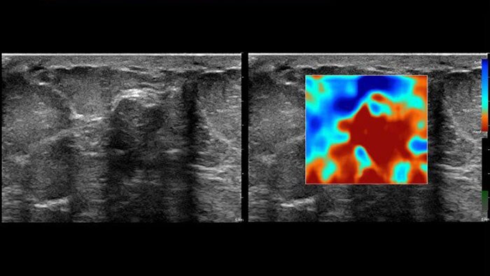
US Q-App Elastography Analysis (EA)*
Explore new tissue stiffness measurements
US Q-App Elastography Analysis (EA)* allows you to strain elastography analysis of tissue deformation based on an elastogram. The applications can be used to size compare between two ROIs; results may be appended to patient reports.
Benefits
- Thumbnail display of frames.
- Size comparison between two ROIs.
- Strain ratio calculation.
- Append to patient exam.
- Compatible with Philips EPIQ, Affiniti and iU22 systems using Elastography mode.
* Only available for sale in the US.
- Q-App Elastography Quantification (EQ)
-
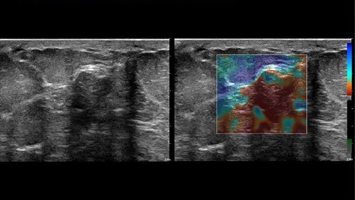
US Q-App Elastography Quantification (EQ)*
Explore new tissue stiffness measurements
US Q-App Elastography Quantification (EQ)* allows you to strain elastography quantification of tissue deformation based on an elastogram. Calculate and display the strain rate and total strain, size compare between two ROIs, and strain ratio; results may be appended to patient reports.
Benefits
- Thumbnail display of frames.
- Size comparison between two ROIs.
- Strain ratio calculation.
- Calculation and display of strain rate and total strain.
- Graphical display of strain ratios with parametric imaging.
- Append to patient exam.
- Compatible with Philips EPIQ , Affiniti and iU22 systems using Elastography mode.
* Not available for sale in the US.
- Q-App General Imaging 3D Quantification
-
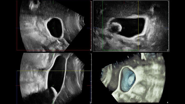
US Q-App General Imaging 3D Quantification (GI3DQ)
Perform advanced visualization and quantification of ultrasound volume
US Q-App General Imaging 3D Quantification (GI3DQ) provides advanced viewing, manipulation, and quantification of 3D data sets. Perform advanced functions such as MPR interrogation, iSlice tomographic imaging, and volume rendering as well as volumetric measurements using multiple methods including semi-automated tools. Results generated from this tool can be appended to the patient’s exam for complete documentation.
Benefits
- Advanced viewing, manipulation, and analysis of 3D ultrasound data.
- Perform multi-planar reconstruction, iSlice tomographic imaging, and advanced rendering functions.
- Easily generate 2D and volume measurements using semi- automated analysis tools.
- Compatible with 3D datasets from Philips EPIQ, Affiniti, iU22, HD15, HD11 and HD9 systems.
- Q-App Imtima Media Thickness (IMT)
-
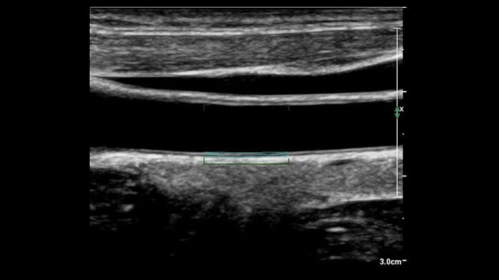
US Q-App Intima Media Thickness (IMT)
Support in determining cardiovascular disease risk
US Q-App Intima Media Thickness (IMT) provides easy and consistent measurement of intima media thickness in carotids and other superficial vessels. Report IMT values and append them to patient reports.
Benefits
- Automated measurement technique on user-selected frames.
- Selector chart to record location and side of vessel where the IMT is measured.
- Quick optimization for thin or thick intima media complexes.
- User-adjustable region of interest.
- User-defined measurement capability.
- Compatible with Philips EPIQ, Affiniti, iE33, iU22, CX50, HD15, HD11, HD7, EnVisor C.0 ultrasound systems.
- Q-App MicroVascular Imaging (MVI)
-
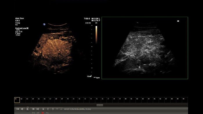
US Q-App MicroVascular Imaging (MVI)
Enhanced vessel conspicuity
US Q-App Microvascular Imaging (MVI) supports you in mapping contrast agent progression with contrast enhanced ultrasound (CEUS) for tumor assessment and monitoring.
Benefits
- Review loops from Philips EPIQ and iU22 systems including side-by-side files.
- Motion compensation algorithm selectable in preferences menu.
- Append to patient exam.
- Compatible with Philips EPIQ, Affiniti , iU22 CX50, and HD15 systems.
- Q-App Region of Interest (ROI)
-
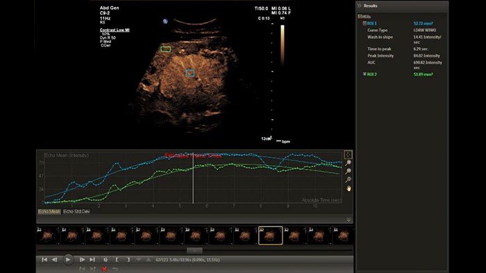
US Q-App Region of Interest (ROI)
Perform advanced analysis of 2D, Color and CEUS data
The Q-App Region of Interest (ROI) provides dedicated tools for spatial and temporal analysis of regions of interest in 2D, color and contrast enhanced* ultrasound exams (CEUS). This Q-App also provides basic 2D measurement tools (distance, area) as well. For CEUS applications, multiple motion compensated regions can be defined for contrast bubble analysis to generate wash-in/wash-out curves for lesion blood flow assessment.
Benefits
- User definable regions for advanced analysis of ultrasound data.
- Allows analysis of echo and color data over time for pixel intensity and distribution curves.
- Supports contrast enhanced* ultrasound exams and facilitates analysis of temporal wash-in/wash-out events.
- Compatible with Philips EPIQ, Affiniti, iE33, iU22, CX50, HD15, HD11 and HD7 systems files.
*Ultrasound contrast agents are approved for use in Left Ventricular Opacification (LVO), focal Liver lesions characterization, and for the evaluation of suspected or known vesicoureteral reflux in pediatric patients’ urinary tract ultrasonography.
- Q-App Vascular Plaque Quantification (VPQ)
-
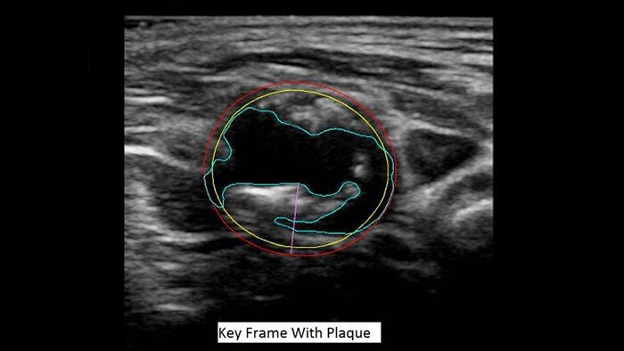
US Q-App Vascular Plaque Quantification (VPQ)
A novel measurement of atherosclerotic plaque volume
US Q-App Vascular Plaque Quantification (VPQ) helps you perform comprehensive volume analysis for carotid plaque; a significant indictor in cardiovascular disease. Automatically measure plaque composition throughout a captured volume, percent area vessel reduction and other characteristics using 3D technology. Results may be posted to patient exams.
Benefits
- 3D technology to visualize and quantify vascular plaque.
- Streamlined workflow through protocol-based task guidance.
- Automatically calculates and displays vessel and plaque boundaries for each frame in the volume data.
- Analysis data presented on image.
- Total plaque volume calculated (mm3).
- Maximum % area reduction calculated.
- Graph analysis data (lumen area, plaque area, reduction over vessel length.
- Compatible with the Philips EPIQ, Affiniti and iU22 systems, monochrome, single-volume 3D volumes acquired with the VL13-5 mechanical transducer.
- Viewing (in MMV)
-
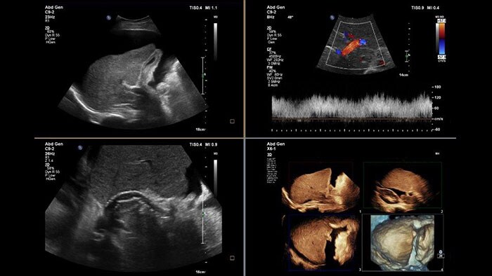
US Viewing (in MMV)
View ultrasound with multi-modality exams on the same workstation
US Viewing (in MMV) and analytics are now available from a multi-modality workstation environment. Review high-resolution single and multi-frame images in collaboration with other modalities. With US Viewing (in Multi Modality Viewer), clinicians can perform measurements, annotations, zoom anatomy and adjust window/levels controls. Edited images can be appended to the patient’s exam for complete documentation. Multi Modality Viewing on IntelliSpace Portal 10 supports additional Q-App tools for advanced quantification of ultrasound data.
Benefits
- View ultrasound single and multi-frame objects.
- User selectable display formats for excellent viewing experience of ultrasound images.
- Easily measure, zoom, perform window-level adjustments and control cine playback functions.
- Append results to exam.